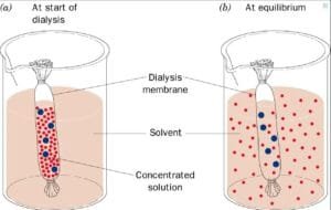
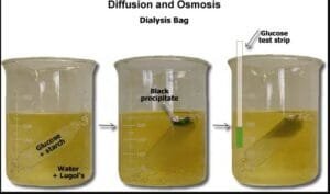
By inspecting the motion of solutes like potassium iodide and glucose, and monitoring weight adjustments in various sucrose concentrations, the experiment goals to exhibit the ideas of passive transport and the way focus gradients impression molecular motion throughout membranes.
This experiment investigates how various concentrations of solutes affect the processes of osmosis and diffusion throughout a semipermeable membrane (dialysis tubing), inspecting the motion of water, solutes, and the ensuing adjustments in resolution traits and weight over time.
Introduction
Osmosis and diffusion are basic processes in mobile transport, taking part in vital roles in sustaining homeostasis and enabling important biochemical features (Alberts et al., 2014). Each are types of passive transport, permitting molecules to maneuver throughout organic membranes with out power. Osmosis is outlined because the motion of water molecules by means of a semipermeable membrane from an space of decrease solute focus to an space of upper solute focus till equilibrium is reached. In distinction, diffusion includes the motion of solute particles from an space of upper focus to an space of decrease focus (Binod, 2024).
Understanding the dynamics of those processes is important for exploring how cells work together with their atmosphere, take up vitamins, and remove waste. There are three primary sorts of options in terms of osmosis: hypertonic, the place there are extra solutes exterior the cell than inside, inflicting water to move out of the cell; hypotonic, the place there are fewer solutes exterior the cell, resulting in water transferring into the cell; and isotonic, the place the focus of solutes is identical inside and out of doors the cell, leading to no internet motion of water (Binod, 2024).
On this experiment, Dialysis luggage are used within the experiment as they’re semipermeable and signify the factitious cells to review the osmosis and diffusion. The speculation of the experiment is that the starch/glucose resolution throughout the dialysis tubing will exhibit a coloration change as a result of diffusion of potassium iodide. And the load of the dialysis luggage will change resulting from osmosis, relying on the sucrose concentrations.
Supplies and Strategies
Steps for Finding out Diffusion and Osmosis Utilizing Dialysis Tubing
Diffusion Experiment:
- Put together the Diffusion Resolution:
- Fill a small beaker with water.
- Add 10 drops of potassium iodide resolution to the beaker to attain a medium brown coloration.
- Observe Diffusion:
- Report the preliminary coloration and look of the potassium iodide resolution within the beaker.
- Look forward to the answer to diffuse, observing the colour change till the answer turns into yellow all through, indicating full diffusion.
- Put together Dialysis Tubing:
- Lower a chunk of dialysis tubing roughly 10 cm lengthy.
- Clamp one finish of the tubing securely and go away the opposite finish open.
- Fill the Dialysis Tubing:
- Fill the open finish of the dialysis tubing about two-thirds full with a starch/glucose resolution.
- Securely clamp the open finish of the tubing.
- Report Preliminary Observations:
- Report the colour of each the potassium iodide resolution within the beaker and the starch/glucose resolution within the dialysis tubing in Desk 1.
- Dip a glucose check strip into the beaker resolution and document the end in Desk 2.
- Place Dialysis Tubing in Beaker:
- Submerge the ready dialysis tubing within the beaker containing the potassium iodide resolution.
- Wait and Observe:
- After half-hour, document the colour adjustments of each the potassium iodide resolution within the beaker and the starch/glucose resolution contained in the dialysis tubing in Desk 1.
- Dip the glucose check strip into the beaker resolution once more and document the end in Desk 2.

Osmosis Experiment:
After 60 minutes, document the ultimate weights of all dialysis cells in Desk 3.
Put together Osmosis Options:
- Fill a small beaker about two-thirds full with a 25% sucrose resolution.
- Fill a big beaker about two-thirds filled with a 1% sucrose resolution.
Put together Dialysis Tubing for Synthetic Cells:
- Get hold of 4 items of soaked dialysis tubing.
- Clip one finish of every piece of dialysis tubing with a clip. Label the clips A, B, C, and D.
Fill Dialysis Tubing:
- Open “Cell A” and fill it about two-thirds full with a 1% sucrose resolution, then securely clamp it.
- Fill “Cell B” with 1% sucrose resolution, “Cell C” with 10% sucrose resolution, and “Cell D” with 25% sucrose resolution, securely clamping every.
Report Preliminary Weights:
- Weigh every of the 4 dialysis cells and document their preliminary weights in Desk 3.
Submerge Dialysis Cells:
- Place “Cell A” within the small beaker with the 25% sucrose resolution.
- Place “Cells B, C, and D” within the massive beaker with the 1% sucrose resolution.
Wait and Weigh:
- After quarter-hour, take away the cells from their respective beakers, dry them barely, and weigh them.
- Report the brand new weight in Desk 3.
- Return the cells to their respective beakers.
Repeat Weighing Course of:
- Repeat the method of eradicating, drying, weighing, and recording the load after half-hour, 45 minutes, and 60 minutes.
Closing Weights:
- After 60 minutes, document the ultimate weights of all dialysis cells in Desk 3.

Outcomes
The outcomes of the experiment exhibit the diffusion of potassium iodide and the conduct of glucose throughout the dialysis tubing, as proven within the following tables.
Desk 1. Diffusion of Potassium iodine resolution from beaker to starch/glucose resolution from Dialysis tubing
| Potassium iodide resolution | Starch/Glucose resolution | |
| Starting coloration | Yellowish brown | white |
| Ending coloration | clear | Purple/cloudy |
Desk 2. Glucose strip check for diffusion
| Colour | Glucose Current? | ||||
| Glucose check strip at starting | teal | damaging | |||
| Glucose check strip at finish | Brown/inexperienced | Hint of glucose | |||
The outcomes of the osmosis experiment are demonstrated by the motion of solutes throughout the dialysis tubing and the adjustments in weight, indicating the hypertonic, hypotonic, or isotonic nature of the atmosphere.
Desk 3. Change in weight of Dialysis cells as a Perform of time
| 0 minutes (preliminary weight) (gm) | 10 minutes weight (gm) | 20 minutes weight (gm) | 30 minutes weight (gm) | 40 minutes weight (gm) | |
| Cell A | 24.16 | 24.53 | 23.90 | 24.12 | 24.65 |
| Cell B | 19.27 | 19.31 | 19.34 | 19.30 | 19.33 |
| Cell C | 24.67 | 24.58 | 24.33 | 24.01 | 23.79 |
| Cell D | 28.50 | 26.87 | 25.65 | 25.57 | 24.78 |
Dialogue
The experiment demonstrated that potassium iodide subtle into the starch/glucose resolution throughout the dialysis tubing, inflicting a coloration change and confirming profitable diffusion. Moreover, the load of the dialysis luggage different primarily based on their surrounding sucrose concentrations, indicating the results of osmosis: cells in hypertonic options misplaced weight, whereas these in isotonic options remained steady.
Preliminary observations revealed that the potassium iodide resolution began as a yellowish brown, whereas the starch/glucose resolution was white. After half-hour, the potassium iodide resolution
grew to become clear, and the starch/glucose resolution turned purple/cloudy, indicating profitable diffusion of the iodine into the tubing.
The glucose check strip initially confirmed a teal coloration, indicating no glucose presence. On the finish of the experiment, the strip modified to brown/inexperienced, confirming a hint of glucose within the beaker resolution.
The burden adjustments over time point out various responses to the encircling options. Cell A confirmed a slight improve in weight after 10 minutes however fluctuated thereafter indicating hypertonic atmosphere within the beaker. Cell B remained comparatively steady indicating an isotonic atmosphere. In distinction, Cell C displayed a gradual lower in weight, whereas Cell D exhibited a major decline, reflecting water loss resulting from its hypertonic atmosphere. The solute strikes from greater focus to decrease focus to keep up homeostasis (Alberts et al., 2014).
The experiment had weaknesses, similar to variations in how properly the dialysis tubing allowed substances to go by means of, which might result in uneven outcomes. Additionally, not controlling the temperature and counting on coloration adjustments for measurements might need made the findings much less dependable.
General, the outcomes assist the speculation that diffusion happens throughout the dialysis membrane and spotlight the results of osmotic stress on the load of the dialysis luggage.
Additional experiments
Future experiments might study at how temperature impacts osmosis and diffusion, anticipating that greater temperatures will make these processes occur sooner. We might additionally strive completely different substances, like salt or sugar, to see how they alter the best way molecules transfer.
References
Alberts, B., Johnson, A., Lewis, J., Raff, M., Roberts, Ok., & Walter, P. (2014). Molecular Biology of the Cell (sixth ed.). Garland Science.
G.C., Binod. “Osmosis and Diffusion: Differences and Factors That Affect Them.” The Science Notes, 14 Apr. 2023. Net. 2 Oct. 2024. Osmosis and Diffusion: Variations and Elements Affecting Them
Mika, T. A., Klein, R. J., Bullerjahn, A. E., Connour, R. L., Swimmer, L. M., White, R. E.,
Gosses, M. W., Carter, T. E., Andrews, A. M., Maier, J. L., & Sidiq, F. (Eds.). (2024). Anatomy and physiology BIO 211 laboratory handbook (third ed.). Owens Group Faculty.
G.C., Binod. “Cellular Transport: Passive and Active Mechanisms.” The Science Notes, 3 Sept. 2024. Net. 2 Oct. 2024. Mobile Transport: Passive and Lively Mechanisms – The Science Notes

